Referral Instructions – Dr. Pyi Naing
Please send consultation and echocardiogram referrals via Medical‑Objects from your clinical software (e.g. Best Practice) using my provider number below. This ensures a secure, efficient, and streamlined referral process.
Medical‑Objects ID / Provider Number: 413245VA
Specialty: Cardiology (General, Heart Failure, Valvular Heart Disease, Pulmonary Hypertension, Dyspnea, Cardiac Risk Assessment and Echocardiography)
Clinic Locations:
Lutwyche: Mandalay Heart Care, Lutwyche HomeCo Shopping Centere, 543 Lutwyche Rd, Lutwyche QLD 4030
Caboolture: Ramsay Specialist Consulting Suites – Caboolture Medical Hub, 124 McKean St, Caboolture QLD 4510
Bribie Island: Ramsay Specialist Consulting Suites – Suite 4, 60 Hornsby Rd, Bongaree QLD 4507
Inpatient Care: St Vincent’s Private Hospital Northside, Chermside QLD 4032
Phone: (07) 2139 8399
Fax: (07) 2145 8015
Email: admin@mandalayheart.com


🫀 GP Echocardiography Referral Guide: Supporting Primary Care Decisions
✅ When to Request an Echocardiogram
Use this guide to support timely, appropriate referrals. Please include clinical context and a clear question.
💔 Suspected Heart Failure
☐ Breathlessness, fatigue, ankle swelling
☐ History of myocardial infarction, hypertension, or cardiomyopathy
📝 Suggested wording: “Assess left ventricular function and valve status in suspected heart failure”
❤️ Heart Murmur or Valve Disease
☐ Murmur noted on examination
☐ Symptoms: exertional dyspnoea, syncope, chest pain
📝 Suggested wording: “Evaluate valve disease severity and haemodynamic impact”
💓 Chest Pain / Post-MI
☐ ECG changes, elevated troponin, or recent myocardial infarction
📝 Suggested wording: “Assess left ventricular function and wall motion post-MI”
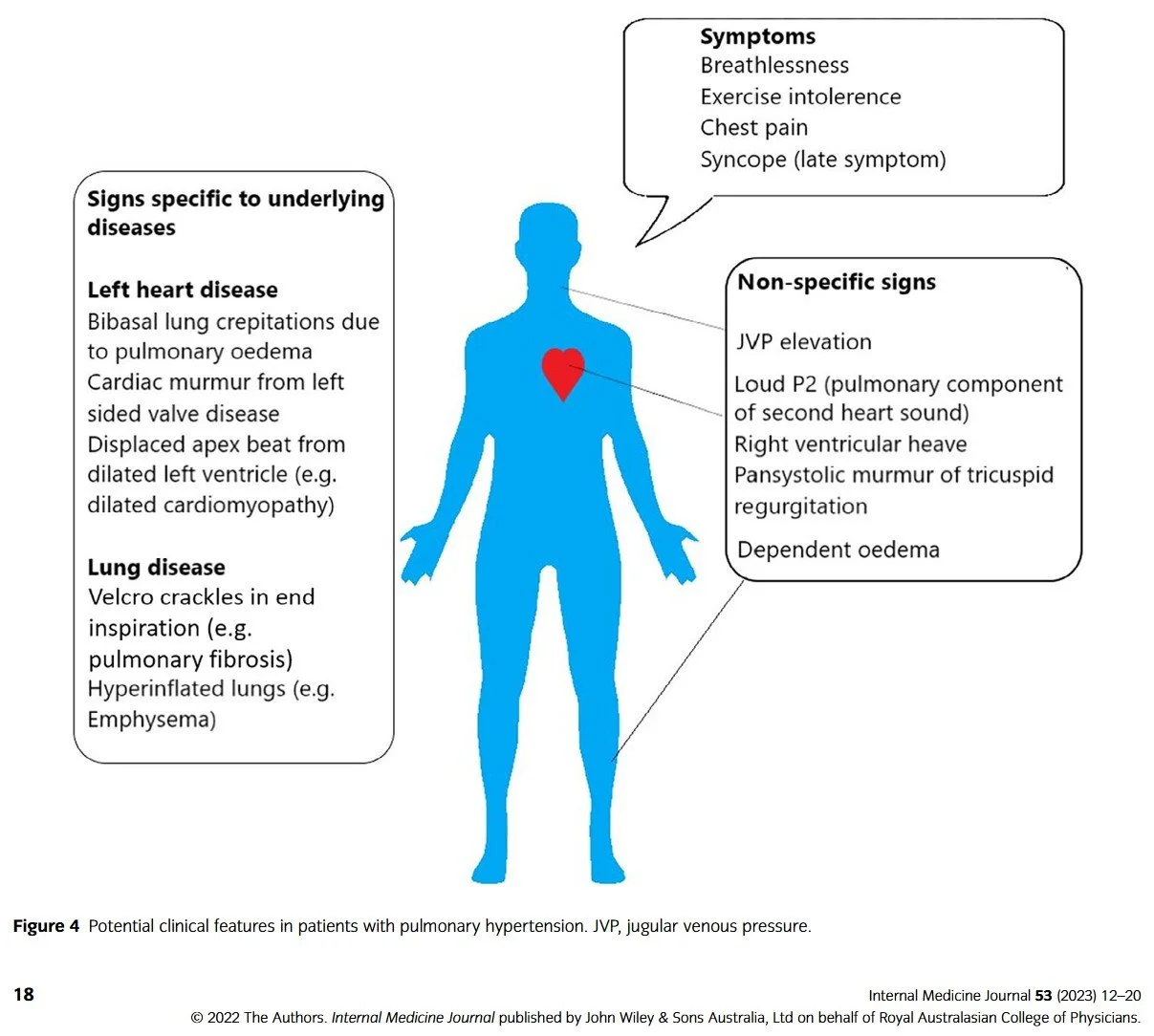
🫁 Pulmonary Hypertension
☐ Unexplained dyspnoea, loud second heart sound, signs of right heart strain
📝 Suggested wording: “Estimate pulmonary pressures and assess right ventricular function”
🫀 Cardiomyopathy Screening
☐ Family history, abnormal ECG, palpitations, syncope
📝 Suggested wording: “Screen for structural heart disease and ventricular function”
🫣 Pericardial Disease
☐ Chest pain, low voltage ECG, raised JVP
📝 Suggested wording: “Assess for pericardial effusion or tamponade physiology”
🧠 Stroke or TIA
☐ Embolic stroke or TIA without clear source
📝 Suggested wording: “Evaluate for cardiac source of embolism (e.g., LV thrombus, PFO)”
🧾 Practical Notes
🔹 Mark as urgent if symptoms are severe or rapidly progressing
🔹 Repeat echo only if there’s a change in clinical status or treatment response
🔹 Consider TOE if poor windows or detailed valve/endocarditis assessment is needed
🔹 Add BNP/NT-proBNP if heart failure diagnosis is uncertain